Lower Forelimb
There are 5 lower forelimb muscles that you will be held accountable for knowing. The extensor carpi ulnaris and radialis, the extensor digitorum lateralis and communis, and the brachioradialis.
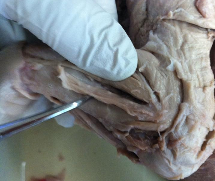
How-To: To find these muscles, look on the lower forelimb for any lines that you can see from the separation of muscles. Use the blunt probe, or if you are confident in your hands, the scissors to cut apart the connective tissue in the forelimb. There is a lot, and it is difficult to remove it all without nicking a muscle or two, but try your best. After removing all the connective tissues, you will have a bunch of parallel lines of muscles, just reference our pictures to know which one is which. from the top of the leg down, it is in this order: brachioradialis, extensor carpi radialis, extensor digitorum communis, extensor digitorum lateralis, extensor carpi ulnaris.
How-To: To find these muscles, look on the lower forelimb for any lines that you can see from the separation of muscles. Use the blunt probe, or if you are confident in your hands, the scissors to cut apart the connective tissue in the forelimb. There is a lot, and it is difficult to remove it all without nicking a muscle or two, but try your best. After removing all the connective tissues, you will have a bunch of parallel lines of muscles, just reference our pictures to know which one is which. from the top of the leg down, it is in this order: brachioradialis, extensor carpi radialis, extensor digitorum communis, extensor digitorum lateralis, extensor carpi ulnaris.
|
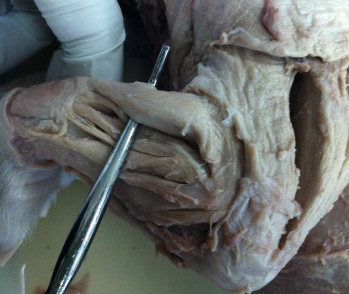
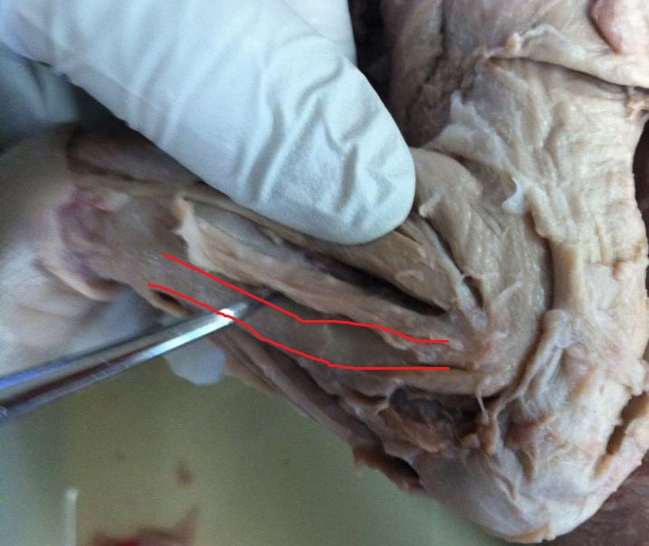
Brachioradialis How-To: The brachioradialis is the most anterior of the lower forelimb muscles. Because it is the outermost and largest of the lower forelimb muscles, it is fairly easy to isolate.
|
|
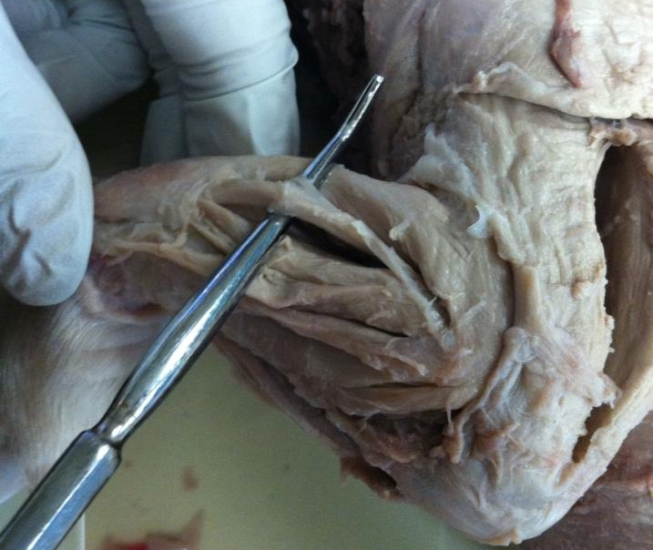
Extensor carpi radialis How-To: The extensor carpi radialis is the muscle that is found immediately deep to the brachioradialis. The radius (bone) is generally under and between the radialis and extensor digitorum communis. We have the probe showing two different lines of muscles, but we seperated more than we had to, the two lines are both extensor carpi radialis. if you see towards the feet, the muscles merge together.
|
|
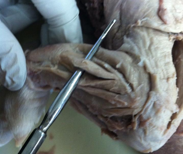
Extensor digitorum communis
How-To: The extensor digitorum communis is the next muscle, after the radialis. This is easily remembered through the word "communis" think of it having two lines communing to make one muscle. this is also easily spotted because it should be the only one that has two seperate divisions in the muscle.
|
|
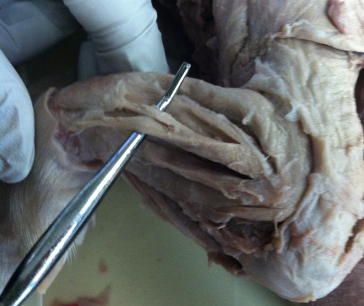
Extensor digitorum lateralis How-To: The lateralis is as the name says, lateral (to the extensor digitorum communis). The lateralis will be the single strand of muscle next to the communis.
|
|
Extensor carpi ulnaris How-To: The ulnaris is the muscle on the last muscle, the most posterior of the 5 forelimb muscles. It is difficult to separate from the ulna because it is tightly connected to the bone. the red line outlines where ulnaris is (not where the probe points to).
|
Forelimb
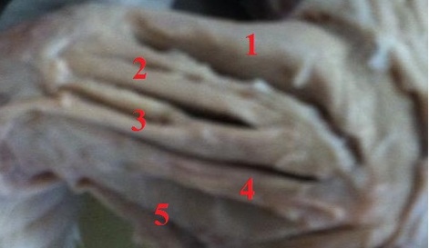
1- brachioradialis
2- extensor carpi radialis (as mentioned above, should not be seperated into two)
3- extensor digitorum communis (should be divided into two)
4- extensor digitorum longus
5- extensor carpi ulnaris
2- extensor carpi radialis (as mentioned above, should not be seperated into two)
3- extensor digitorum communis (should be divided into two)
4- extensor digitorum longus
5- extensor carpi ulnaris
