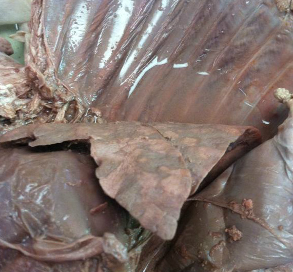
Above, you can see the left lung in the foreground of the picture. You can see discolorations in the lung, the white spots are the bronchioles which are clustered about in the lung (scrape the lung some and more and more white material will show up, these are all bronchioles).
Bronchi
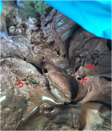
The picture shows the medial sides of the lungs. the arrows point to where the bronchi go in the lungs, making little holes of opening on the lung surface.
To find this, you must open the thoracic cavity and remove the heart.
To find this, you must open the thoracic cavity and remove the heart.
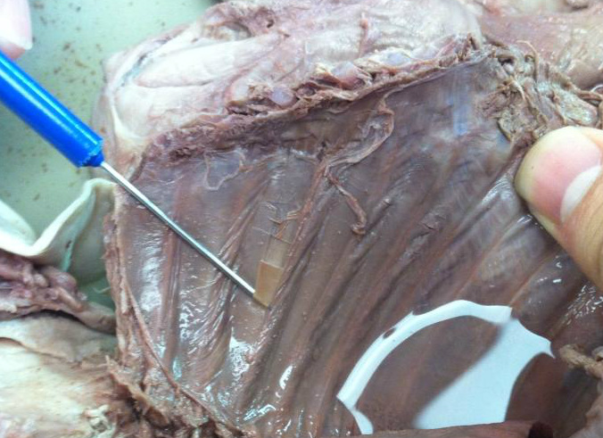
The parietal pleura is a serous membrane which surrounds the lungs, is shown being lifted up by the straight teasing needle. It is like a thin film, and can be somewhat difficult to remove and isolate. the parietal pleura is shown on the thoracic cavity wall in this picture because it was attached to the wall and ripped from the lung surface. the Visceral pleura is seen on the layer underneath the thin membrane held up by the needle.
in our
in our
